Healthy Lung X Ray Side View

Basal lung consolidation. this image shows subtle consolidation at the left lung base, partly obscured by the heart; if you are in doubt about a certain appearance on an x-ray, make sure you check to see if the patient has had previous images see next image. Smoking is the leading cause of lung cancer. even if you dont smoke, being around someone who smokes on a regular basis and inhaling their secondhand smoke can also cause cancer. when you inhale cigarette smoke, the carcinogens cause changes in the tissues and cells in the lungs. over time, these changes damage the genetic material of the cells in the lungs and cause cancer to develop. a healthy lung and one damaged by smoking look very different. a lung damaged by smoking is blackened over time, and its shape becomes irregular and hardened.

Chest Xraynormal Youtube

step stool (double) medical kick bucket hospital bed side screen view all x-ray view box (cfl tube illumination) seating stools medical baby bassinet (with utility box) hospital baby bassinet x-ray view box (led illumination) hospital bed side screen code : mf4201 medical kick bucket code : mf4301 An x-ray from this view is taken with the patient lying down on the side. it helps to assess suspected fluid (pleural effusion), and demonstrate whether the effusion is loculated or mobile. you can look at the non-dependent hemithorax to confirm a pneumothorax. the dependant lung should increase in density.
Chest Xray For The Diagnosis Of Lung Cancer Verywell Health
The chest x-ray is one of the most common imaging tests performed in clinical practice, typically for cough, shortness of breath, chest pain, chest wall trauma, and assessment for occult disease. standard x-rays are performed with the patient standing facing an x-ray film or digital cassette, 6 feet away from an x-ray healthy lung x ray side view tube. Chestx-ray views. the x-ray of chest is may be taken from different angles based on the direction of passing the ionizing x-rays. it is referred to as ‘views’. posterior-anterior (pa) view refers to x-ray images taken by allowing x-rays to pass from the back side of the body to the front side of chest and fall on the x-ray film placed in.
An x-ray from this view is taken with the patient lying down on the side. it helps to assess suspected fluid (pleural effusion), and demonstrate whether the effusion is loculated or mobile. you can look at the non-dependent hemithorax to confirm a pneumothorax.
Chest Xraynormal Youtube
Lung zones. assess the lungs by comparing the upper, middle and lower lung zones on the left and right. asymmetry of lung density is represented as either abnormal whiteness (increased density), or abnormal blackness (decreased density). once you have spotted asymmetry, the next step is to decide which side is abnormal. Chest x-rays are a common type of exam. a chest x-ray is often among the first procedures you'll undergo if your doctor suspects you have heart or lung disease. it can also be used to check how you are responding to treatment. a chest x-ray can reveal many things inside your body, including: 1. the condition of your lungs. chest x-rays can detect cancer, infection or air collecting in the space around a lung (pneumothorax). they can also show chronic lung conditions, such as emphysema or cysti Looking at this in a different way, a 2013 review of radiology malpractice suits involving the thorax (the chest cavity), found that at least 40 percent of cases were related to a missed diagnosis of lung cancer. anatomically, cancers in certain parts of the lungs are more difficult to visualize and are more likely to be missed on a chest x-ray. as noted earlier, dense structures such as bone can \\"hide\\" small cancers. in fact, in one study, 72 percent of missed lung cancers were in the upper lobes and of these 22 percent were obscured by the collar bones (clavicles. ) cancers found in the periphery of the lungs (such as lung adenocarcinoma) are more commonly missed than those that occur centrally near the large airways (such as small cell lung cancer and squamous cell carcinoma of the lungs. ).
Chest Xray Pulmonary Disease Normal Comparison
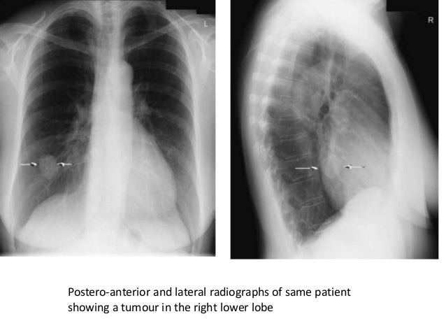
See full list on healthline. com. A chest x-ray helps detect problems with your heart and lungs. the chest x-ray on the left is normal. the image on the right shows a mass in the right lung. See full list on verywellhealth. com. A chest x-ray helps detect problems with your heart and lungs. the chest x-ray on the left is normal. the image on the right shows a mass in the right lung.
Plain chest x-rays miss a diagnosis of lung cancer far too often. there are surprisingly few recent studies looking at the actual incidence of \\"missed diagnoses\\" of lung cancer, but the research that has been done is sobering. it can be frightening to hear about the incidence of missed lung cancers on chest x-rays, but this doesn't mean you need despair. as noted above, ct screening can decrease the risk of death from lung cancer among those who have risk factors. yet even for those without these risk factors, there are several things you can do: chest x-rays may be helpful in finding a lung cancer but cannot exclude the presence of cancer. in contrast, a normal chest x-ray may convey the false reassurance that everything is okay. chest x-rays can miss small and potentially curable lung cancers. if you have unexplained symptoms or risk factors for lung cancer, make sure you talk to your doctor. There are several different tests that will allow your doctor to make a lung cancer diagnosis: if you have any symptoms of lung cancer, your doctor may order a chest x-ray. a chest x-ray of someone with lung cancer may show a visible mass or nodule. this mass will look like a white spot on your lungs, while the lung itself will appear black. however, an x-ray may not be able to detect small or early-stage cancers. a computed tomography (ct) scan is often ordered if there is something abnormal on the chest x-ray. a ct scan takes a cross-sectional and a more detailed image of the lung. it can give more information about any abnormalities, nodules, or lesions small, abnormal areas in the lungs that were seen on x-ray. a ct scan can detect smaller lesions that are not visible on a chest x-ray. cancerous lesions can often be distinguished from benign lesions on chest ct scans. your doctor cannot give you a cancer diagnosis with only an image from a ct scan or an x-ray. if they are concerned about the results of your image tests they will order a tissue biopsy. in a biopsy, your physician will take a tissue sample from your lungs for examination. this sample may be removed via a tube placed down your throat (bronchoscopy) or by making an incision in the chest wall and using a needle to collect the sample. this sample can then be analyzed by a pathologist to determine if you have cancer. Mar 29, 2019 · an x-ray from this view is taken with the patient lying down on the side. it helps to assess suspected fluid (pleural effusion), and demonstrate whether the effusion is loculated or mobile. you can look at the non-dependent hemithorax to confirm a pneumothorax. the dependant lung should increase in density.
Chest x-ray for the diagnosis of lung cancer verywell health.
24, 2012 september 17, 2013 author admin categories healthy eating tags articles cooking eating fastfood food health mcdonald nutrition unhealthy eating leave a comment on unhealthy eating lung cancer x-ray image via wikipedia get to know more about Nov 12, 2018 · a chest x-ray is one method of providing your doctor with images of your heart and lungs. a computed tomography (ct) scan of the chest is another tool that is commonly ordered in people with. For those who have symptoms of lung cancer, a large 2006 study in the u. k. found that almost 25 percent of chest x-rays done in the primary care setting for patients with lung cancer were negative when performed within a year of diagnosis. negative chest x-rays occurred in people with all of the most common symptoms of healthy lung x ray side view lung cancer with the exception of hoarseness.
A word from verywell. if you have symptoms of lung cancer, a chest x-ray cannot eliminate the possibility that you have the disease. as reassuring as a "normal" result may seem, don't allow it to give you a false sense of security if the cause of persistent symptoms remains unknown or if the diagnosis you were given can't explain them. this is even true for never-smokers in whom lung cancer. In 2016, 224,390 people in the united states will be diagnosed with lung cancer. the diagnosis of lung cancer is very healthy lung x ray side view serious. lung cancer kills more people than colon, breast, and prostate cancer combined. it is more common in men than in women, and african american men are 20 percent more likely than caucasian men to have lung cancer. early diagnosis and treatment are important for survival.

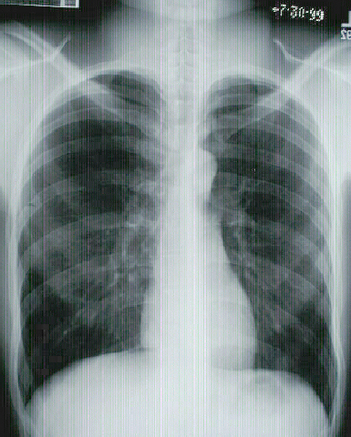
Normal chest x-ray. please see disclaimer on healthy lung x ray side view my website. www. my-uni. net. The lungs are a pair of spongy, air-filled organs located on either side of the chest (thorax). the trachea (windpipe) conducts inhaled air into the lungs through its tubular branches, called bronchi. the bronchi then divide into smaller and smaller branches (bronchioles), finally becoming microscopic. the bronchioles eventually end in clusters of microscopic air sacs called alveoli. A word from verywell. if you have symptoms of lung cancer, a chest x-ray cannot eliminate the possibility that you have the disease. as reassuring as a "normal" result may seem, don't allow it to give you a false sense of security if the cause of persistent symptoms remains unknown or if the diagnosis you were given can't explain them. See full list on mayoclinic. org.
Komentar
Posting Komentar